Copyright
©2005 Baishideng Publishing Group Inc.
World J Gastroenterol. Feb 21, 2005; 11(7): 1052-1055
Published online Feb 21, 2005. doi: 10.3748/wjg.v11.i7.1052
Published online Feb 21, 2005. doi: 10.3748/wjg.v11.i7.1052
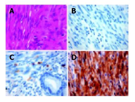
Figure 1 Light microscopic examination of case #23 (A) shows interlacing spindle tumor cells (hematoxylin & eosin stain).
The tumor cells were negative for CD117 by immunohistochemistry (B), while the adjacent mast cells were immunoreative to CD117 (C). This tumor was strongly positive for S-100 (D). (magnification ×400).
- Citation: Tzen CY, Mau BL. Analysis of CD117-negative gastrointestinal stromal tumors. World J Gastroenterol 2005; 11(7): 1052-1055
- URL: https://www.wjgnet.com/1007-9327/full/v11/i7/1052.htm
- DOI: https://dx.doi.org/10.3748/wjg.v11.i7.1052


