Copyright
©2005 Baishideng Publishing Group Inc.
World J Gastroenterol. Feb 14, 2005; 11(6): 880-884
Published online Feb 14, 2005. doi: 10.3748/wjg.v11.i6.880
Published online Feb 14, 2005. doi: 10.3748/wjg.v11.i6.880
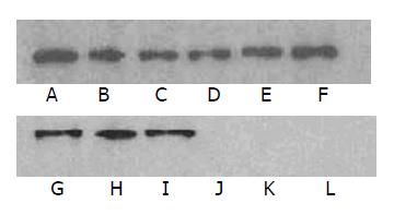
Figure 2 Tyrosine phosphorylation sites in EPIYA in vitro and in vivo.
A-F: tyrosine phosphorylation in vitro. Lane A: CagA with 4 EPIYAs; lane B: CagA with 3 EPIYAs; lane C: CagA with 2 EPIYAs; lane D: EPIFA with 1 EPIYA; lane E: EPIFA with 2 EPIYAs; lane F: EPIFA with 3 EPIYAs. lane G-L: tyrosine phosphorylation in vivo. G: CagA with 4 EPIYAs; H: CagA with 3 EPIYAs; I: CagA with 2 EPIYAs; lane J: EPIFA with 3 EPIYAs; lane K: EPIFA with 2 EPIYAs; lane L: EPIFA with 1 EPIYA.
-
Citation: Zhu YL, Zheng S, Du Q, Qian KD, Fang PC. Characterization of
CagA variable region ofHelicobacter pylori isolates from Chinese patients. World J Gastroenterol 2005; 11(6): 880-884 - URL: https://www.wjgnet.com/1007-9327/full/v11/i6/880.htm
- DOI: https://dx.doi.org/10.3748/wjg.v11.i6.880


