Copyright
©2005 Baishideng Publishing Group Inc.
World J Gastroenterol. Dec 28, 2005; 11(48): 7694-7696
Published online Dec 28, 2005. doi: 10.3748/wjg.v11.i48.7694
Published online Dec 28, 2005. doi: 10.3748/wjg.v11.i48.7694
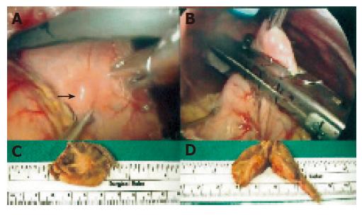
Figure 3 Localization of the tumor mass by intra-operative upper tract endoscopy.
A: Tumor lesion in the posterior wall of gastric high body; B: robotic-assisted laparoscopic wedge resection of the gastric high body; C: submucosal tumor with tumor-free margin; D: cutting surface with firm, yellow, well-circumscribed, lobulated nodules.
- Citation: Hsu SD, Wu HS, Kuo CL, Lee YT. Robotic-assisted laparoscopic resection of ectopic pancreas in the posterior wall of gastric high body: Case report and review of the literature. World J Gastroenterol 2005; 11(48): 7694-7696
- URL: https://www.wjgnet.com/1007-9327/full/v11/i48/7694.htm
- DOI: https://dx.doi.org/10.3748/wjg.v11.i48.7694


