Copyright
©2005 Baishideng Publishing Group Inc.
World J Gastroenterol. Dec 28, 2005; 11(48): 7690-7693
Published online Dec 28, 2005. doi: 10.3748/wjg.v11.i48.7690
Published online Dec 28, 2005. doi: 10.3748/wjg.v11.i48.7690
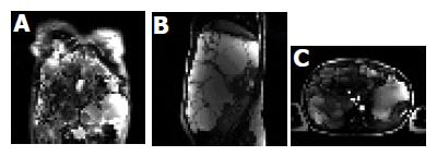
Figure 1 Massive polycystic liver disease on coronal MR imaging (A), caudal and posterior displacement of abdominal organs by the massively enlarged liver on sagittal MR imaging (B), and compression of the inferior vena cava (arrow) produced by the massively enlarged liver (C) on MR angiograph.
- Citation: Peces R, Drenth JP, Morsche RHT, González P, Peces C. Autosomal dominant polycystic liver disease in a family without polycystic kidney disease associated with a novel missense protein kinase C substrate 80K-H mutation. World J Gastroenterol 2005; 11(48): 7690-7693
- URL: https://www.wjgnet.com/1007-9327/full/v11/i48/7690.htm
- DOI: https://dx.doi.org/10.3748/wjg.v11.i48.7690


