Copyright
©2005 Baishideng Publishing Group Inc.
World J Gastroenterol. Dec 28, 2005; 11(48): 7620-7624
Published online Dec 28, 2005. doi: 10.3748/wjg.v11.i48.7620
Published online Dec 28, 2005. doi: 10.3748/wjg.v11.i48.7620
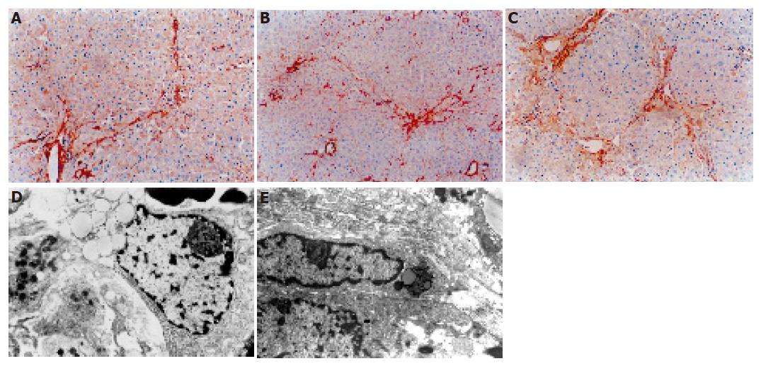
Figure 4 Distribution of α-SMA positive cells after DMN treatment.
A: After 14 d, linear immunoreaction for α-SMA was scattered along the sinusoidal wall; B: after 21 d, a network of α-SMA cells was evident; C: after 28 d, a dense network of α-SMA cells was evident; D: after 14 d, transitional hepatic stellate cells were observed under electron microscope; E: after 21 or 28 d, typical myofibroblasts were embedded in the core of fibrous septa.
- Citation: Li CH, Piao DM, Xu WX, Yin ZR, Jin JS, Shen ZS. Morphological and serum hyaluronic acid, laminin and type IV collagen changes in dimethylnitrosamine-induced hepatic fibrosis of rats. World J Gastroenterol 2005; 11(48): 7620-7624
- URL: https://www.wjgnet.com/1007-9327/full/v11/i48/7620.htm
- DOI: https://dx.doi.org/10.3748/wjg.v11.i48.7620


