Copyright
©2005 Baishideng Publishing Group Inc.
World J Gastroenterol. Dec 14, 2005; 11(46): 7284-7289
Published online Dec 14, 2005. doi: 10.3748/wjg.v11.i46.7284
Published online Dec 14, 2005. doi: 10.3748/wjg.v11.i46.7284
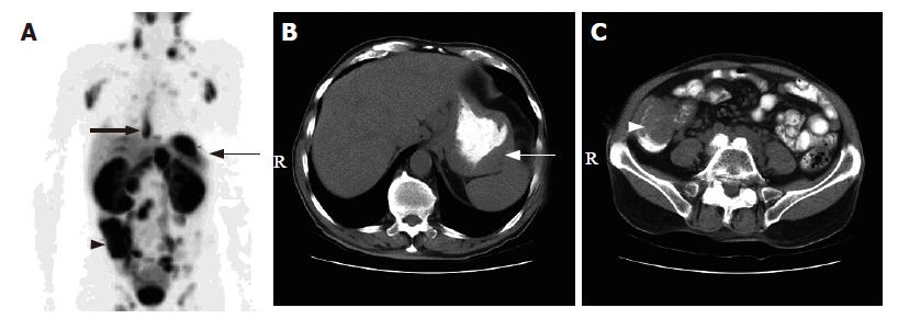
Figure 4 A 78-year-old man with mantle cell lymphoma.
A: 18F-FDG PET scan revealed multiple hypermetabolic foci involving the lower esophagus (thick arrow), gastric fundus (thin arrow), cecum and terminal ileum (arrow head); B and C: The corresponding CT scans of the abdomen showed wall thickening at the gastric fundus (white arrow) and the cecum and terminal ileum (white arrow head)
- Citation: Phongkitkarun S, Varavithya V, Kazama T, Faria SC, Mar MV, Podoloff DA, Macapinlac HA. Lymphomatous involvement of gastrointestinal tract: Evaluation by positron emission tomography with 18F-fluorodeoxyglucose. World J Gastroenterol 2005; 11(46): 7284-7289
- URL: https://www.wjgnet.com/1007-9327/full/v11/i46/7284.htm
- DOI: https://dx.doi.org/10.3748/wjg.v11.i46.7284


