Copyright
©2005 Baishideng Publishing Group Inc.
World J Gastroenterol. Dec 14, 2005; 11(46): 7284-7289
Published online Dec 14, 2005. doi: 10.3748/wjg.v11.i46.7284
Published online Dec 14, 2005. doi: 10.3748/wjg.v11.i46.7284
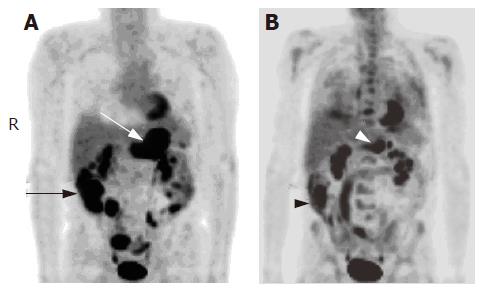
Figure 2 A 68-year-old man with large cell lymphoma.
A: Base line 18F-FDG PET scan showed hypermetabolic foci in the stomach (white arrow), cecum and terminal ileum (black arrow), and bowel loops (SUVmax = 12.7); B: 18F-FDG PET scan obtained after therapy showed partial metabolic response of the activity in the stomach (white arrowhead) and cecum (black arrowhead, SUVmax = 10.7).
- Citation: Phongkitkarun S, Varavithya V, Kazama T, Faria SC, Mar MV, Podoloff DA, Macapinlac HA. Lymphomatous involvement of gastrointestinal tract: Evaluation by positron emission tomography with 18F-fluorodeoxyglucose. World J Gastroenterol 2005; 11(46): 7284-7289
- URL: https://www.wjgnet.com/1007-9327/full/v11/i46/7284.htm
- DOI: https://dx.doi.org/10.3748/wjg.v11.i46.7284


