Copyright
©2005 Baishideng Publishing Group Inc.
World J Gastroenterol. Dec 7, 2005; 11(45): 7063-7071
Published online Dec 7, 2005. doi: 10.3748/wjg.v11.i45.7063
Published online Dec 7, 2005. doi: 10.3748/wjg.v11.i45.7063
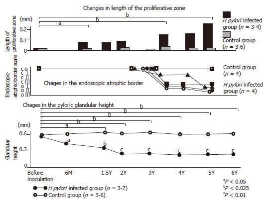
Figure 1 Gastric mucosal alteration of Japanese monkey model with H pylori infection.
Upper graph showed the gradual increase of the proliferative zone of H pylori-infected Japanese monkey model. Middle graph showed the alteration of endoscopic-atrophic-border scale of this model. Macroscopically, gastric atrophy advanced for more than 3 yr. Lower graph showed the alteration of the pyloric glandular height. Six months after inoculation, the pyloric glandular height was apparently lower in the infected animals than in controls. Furthermore, the atrophic change advanced gradually throughout the 6-yr observation period.
- Citation: Kodama M, Murakami K, Sato R, Okimoto T, Nishizono A, Fujioka T. Helicobacter pylori-infected animal models are extremely suitable for the investigation of gastric carcinogenesis. World J Gastroenterol 2005; 11(45): 7063-7071
- URL: https://www.wjgnet.com/1007-9327/full/v11/i45/7063.htm
- DOI: https://dx.doi.org/10.3748/wjg.v11.i45.7063


