Copyright
©2005 Baishideng Publishing Group Inc.
World J Gastroenterol. Nov 28, 2005; 11(44): 7028-7032
Published online Nov 28, 2005. doi: 10.3748/wjg.v11.i44.7028
Published online Nov 28, 2005. doi: 10.3748/wjg.v11.i44.7028
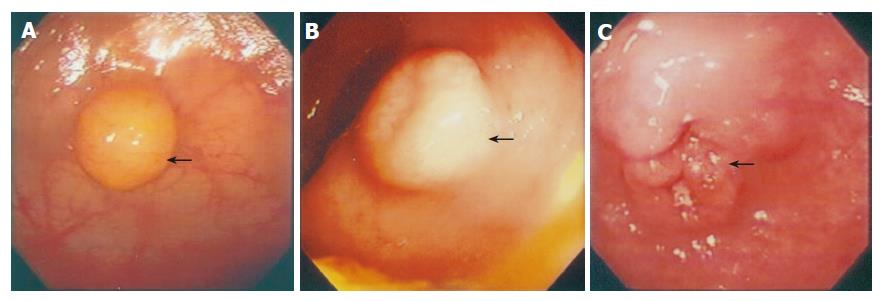
Figure 2 Subject A: A polypoid carcinoid lesion at the gastric fundus site about 0.
8 cm with typical yellowish color of the lesion; B: A submucosal carcinoid lesion at the duodenal bulb about 1.7 cm; C: A carcinoid tumor with ulceration, at the antrum site, about 4.0 cm in size.
- Citation: Chuah SK, Hu TH, Kuo CM, Chiu KW, Kuo CH, Wu KL, Chou YP, Lu SN, Chiou SS, Changchien CS, Eng HL. Upper gastrointestinal carcinoid tumors incidentally found by endoscopic examinations. World J Gastroenterol 2005; 11(44): 7028-7032
- URL: https://www.wjgnet.com/1007-9327/full/v11/i44/7028.htm
- DOI: https://dx.doi.org/10.3748/wjg.v11.i44.7028


