Copyright
©2005 Baishideng Publishing Group Inc.
World J Gastroenterol. Nov 28, 2005; 11(44): 6932-6935
Published online Nov 28, 2005. doi: 10.3748/wjg.v11.i44.6932
Published online Nov 28, 2005. doi: 10.3748/wjg.v11.i44.6932
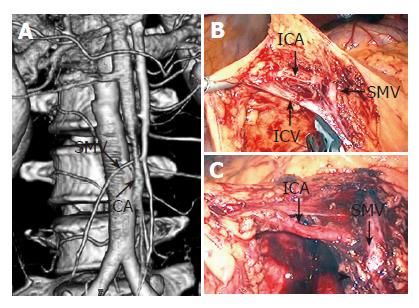
Figure 2 ICA situated in back of SMV at 3DCT (A) and at intraoperative findings (B); C: ICA existed after the ileocecal vein (ICV) had been resected.
The arrow head shows the cut end of the ICV.
- Citation: Ohtani H, Ohta K, Arimoto Y, Kim EC, Oba H, Adachi K, Terakawa S, Tsubakimoto M. Three-dimensional computed tomography in laparoscopic surgery for colorectal carcinoma. World J Gastroenterol 2005; 11(44): 6932-6935
- URL: https://www.wjgnet.com/1007-9327/full/v11/i44/6932.htm
- DOI: https://dx.doi.org/10.3748/wjg.v11.i44.6932


