Copyright
©2005 Baishideng Publishing Group Inc.
World J Gastroenterol. Nov 28, 2005; 11(44): 6926-6931
Published online Nov 28, 2005. doi: 10.3748/wjg.v11.i44.6926
Published online Nov 28, 2005. doi: 10.3748/wjg.v11.i44.6926
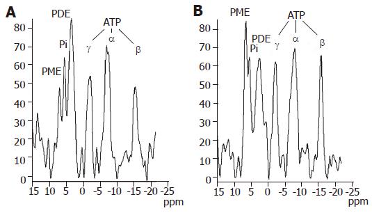
Figure 2 31P MR spectra of the liver in a healthy volunteer (A) and in a patient with liver cirrhosis (B).
PME - phosphomonoesters; Pi - inorganic phosphate; PDE - phosphodiesters; γATP, αATP, βATP - γ, α and β phosphates of adenosine triphosphate.
- Citation: Dezortova M, Taimr P, Skoch A, Spicak J, Hajek M. Etiology and functional status of liver cirrhosis by 31P MR spectroscopy. World J Gastroenterol 2005; 11(44): 6926-6931
- URL: https://www.wjgnet.com/1007-9327/full/v11/i44/6926.htm
- DOI: https://dx.doi.org/10.3748/wjg.v11.i44.6926


