Copyright
©2005 Baishideng Publishing Group Inc.
World J Gastroenterol. Nov 21, 2005; 11(43): 6765-6769
Published online Nov 21, 2005. doi: 10.3748/wjg.v11.i43.6765
Published online Nov 21, 2005. doi: 10.3748/wjg.v11.i43.6765
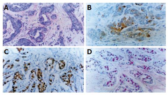
Figure 3 H&E and immunohistochemical staining of pancreatic cancer.
A: Moderate to poorly differentiated pancreatic adenocarcinoma with desmoplastic background. H&E stain, ×200; B: Immunohistochemical staining using CEA monoclonal antibody shows positive reaction in the cytoplasms of tumor cells. DAB and hematoxylin counter stain, ×200; C: Ki-67 monoclonal antibody shows significantly increased proliferation of nuclei of tumor cells. DAB and hematoxylin counter stain, ×200; D: Intense nuclear immunohistochemical staining of p53 in an invasive pancreatic adenocarcinoma. AEC and hematoxylin counter stain, ×200.
-
Citation: Jeong S, Lee DH, Lee JI, Lee JW, Kwon KS, Kim PS, Kim HG, Shin YW, Kim YS, Kim YB. Expression of Ki-67, p53, and K-
ras in chronic pancreatitis and pancreatic ductal adenocarcinoma. World J Gastroenterol 2005; 11(43): 6765-6769 - URL: https://www.wjgnet.com/1007-9327/full/v11/i43/6765.htm
- DOI: https://dx.doi.org/10.3748/wjg.v11.i43.6765


