Copyright
©The Author(s) 2005.
World J Gastroenterol. Nov 14, 2005; 11(42): 6694-6700
Published online Nov 14, 2005. doi: 10.3748/wjg.v11.i42.6694
Published online Nov 14, 2005. doi: 10.3748/wjg.v11.i42.6694
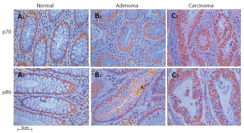
Figure 5 Expression of DNA-PK protein subunits in human colon tissues.
Tissue sections were stained by immunohistochemistry for Ku70 and Ku86 proteins, as described in “Materials and methods”. The increase of positively stained cells is evident in the nuclei of adenoma and carcinoma sections, respect to the normal controls. Ku70 nuclear expression is uniform and displays a higher intensity of staining as compared with p86, as described in the text. No cytoplasmic expression is evident in the normal mucosa sections for any of the protein subunits, whereas it is clearly visible in the pathologic tissues. Ku86 was expressed in the cytoplasm following a speckled pattern of staining, as evident on the adenoma section (arrow). Original magnification ×400. A1-2: Normal; B1-2: Adenoma; C1-2: Carcinoma.
- Citation: Mazzarelli P, Parrella P, Seripa D, Signori E, Perrone G, Rabitti C, Borzomati D, Gabbrielli A, Matera MG, Gravina C, Caricato M, Poeta ML, Rinaldi M, Valeri S, Coppola R, Fazio VM. DNA end binding activity and Ku70/80 heterodimer expression in human colorectal tumor. World J Gastroenterol 2005; 11(42): 6694-6700
- URL: https://www.wjgnet.com/1007-9327/full/v11/i42/6694.htm
- DOI: https://dx.doi.org/10.3748/wjg.v11.i42.6694


