Copyright
©2005 Baishideng Publishing Group Inc.
World J Gastroenterol. Nov 14, 2005; 11(42): 6631-6637
Published online Nov 14, 2005. doi: 10.3748/wjg.v11.i42.6631
Published online Nov 14, 2005. doi: 10.3748/wjg.v11.i42.6631
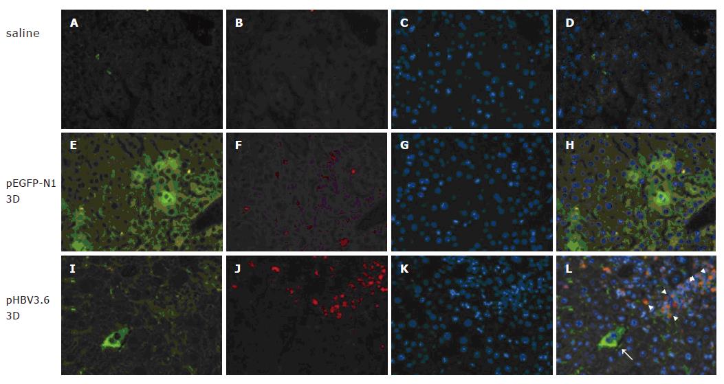
Figure 1 TLR4 expression in the liver of C3H/HeN mice after hydro-dynamic injection of pHBV3.
6. Groups of four C3H/HeN mice were injected intravenously with 10 μg of plasmid by hydrodynamics-based transfection. The liver tissues were collected at day 3 post injection and 4-μm cryosections were made, stained with anti-HBsAg-FITC and anti-TLR4-PE antibody. The green represents EGFP-positive (E) or HBsAg-positive cells (I) and the red represents TLR4-positive cells (B, F, J). The blue represents counter staining by Hoechst 33258 dye (C, G, K). A-D: naïve; E-H: pEGFP-N1; I–L: pHBV3.6. The arrows indicate the HBsAg positive hepatocytes and the arrowheads indicate the TLR4 positive immune cells (original magnification ×200)
- Citation: Chang WW, Su IJ, Lai MD, Chang WT, Huang W, Lei HY. Toll-like receptor 4 plays an anti-HBV role in a murine model of acute hepatitis B virus expression. World J Gastroenterol 2005; 11(42): 6631-6637
- URL: https://www.wjgnet.com/1007-9327/full/v11/i42/6631.htm
- DOI: https://dx.doi.org/10.3748/wjg.v11.i42.6631


