Copyright
©2005 Baishideng Publishing Group Inc.
World J Gastroenterol. Nov 7, 2005; 11(41): 6554-6556
Published online Nov 7, 2005. doi: 10.3748/wjg.v11.i41.6554
Published online Nov 7, 2005. doi: 10.3748/wjg.v11.i41.6554
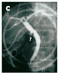
Figure 1 Endoscopic retrograde cholangiography.
The cystic duct is poorly opacified with small protruding lesions around the confluence of the cystic duct (arrow). An 8-Fr pig-tail catheter (C) is inserted into the gallbladder.
- Citation: Wakai T, Shirai Y, Hatakeyama K. Peroral cholangioscopy for non-invasive papillary cholangiocarcinoma with extensive superficial ductal spread. World J Gastroenterol 2005; 11(41): 6554-6556
- URL: https://www.wjgnet.com/1007-9327/full/v11/i41/6554.htm
- DOI: https://dx.doi.org/10.3748/wjg.v11.i41.6554


