Copyright
©2005 Baishideng Publishing Group Inc.
World J Gastroenterol. Oct 28, 2005; 11(40): 6373-6380
Published online Oct 28, 2005. doi: 10.3748/wjg.v11.i40.6373
Published online Oct 28, 2005. doi: 10.3748/wjg.v11.i40.6373
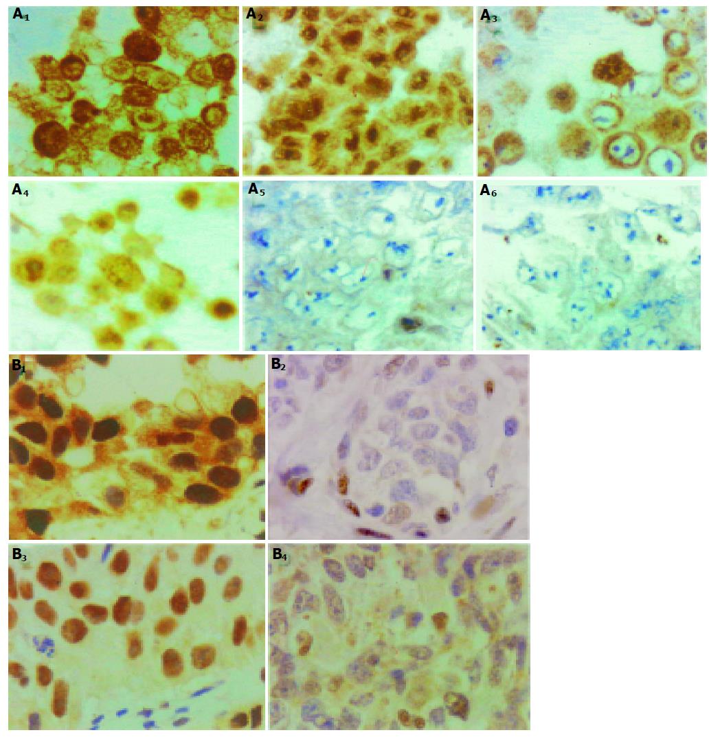
Figure 4 A: Immunocytochemical stain for GR expression in representing carcinoma cell lines.
(A1) and A2): SiHa cells, which had GR content about 8.1×10⁴/cell according to ligand binding assay; (A3) and (A4): HeLa cells, with GR content about 1.97×10⁴/cell; (A5) and (A6): N87 cells, with GR content about 5.0×10³/cell. (A2), (A4), and (A5): cells treated with DEX for 3 h before harvest. In (A1) and (A3), the GR immunoreactivity localized in cytoplasm. After DEX treatment, the immunoreactive GR translocalized to nuclei (A2) and (A4). The low GR content cancer cells N87 showed negligible immunoreactivity. B: Immunohistochemical stain for GR expression in human carcinoma tumor tissue samples. Non-small cell lung cancer tumor samples, with high GR expression (Ba), and low GR expression (B2). Breast cancer tumor samples, with high GR expression (B3), and low GR expression (B4).
- Citation: Lu YS, Lien HC, Yeh PY, Yeh KH, Kuo ML, Kuo SH, Cheng AL. Effects of glucocorticoids on the growth and chemosensitivity of carcinoma cells are heterogeneous and require high concentration of functional glucocorticoid receptors. World J Gastroenterol 2005; 11(40): 6373-6380
- URL: https://www.wjgnet.com/1007-9327/full/v11/i40/6373.htm
- DOI: https://dx.doi.org/10.3748/wjg.v11.i40.6373


