Copyright
©2005 Baishideng Publishing Group Inc.
World J Gastroenterol. Oct 28, 2005; 11(40): 6354-6359
Published online Oct 28, 2005. doi: 10.3748/wjg.v11.i40.6354
Published online Oct 28, 2005. doi: 10.3748/wjg.v11.i40.6354
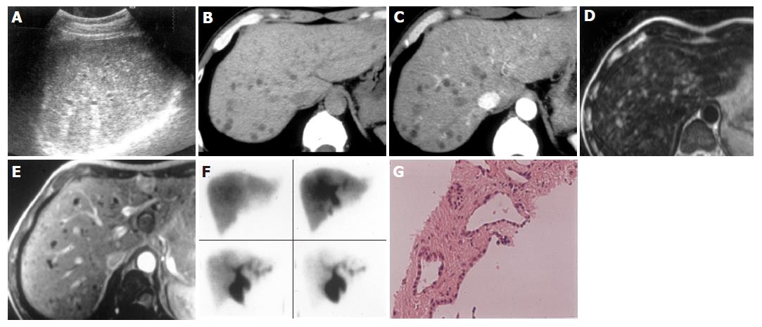
Figure 1 A 51-year-old man with VMCs.
A: B-mode US showed multiple small hyper- and hypoechoic lesions less than 10 mm in diameter with multiple comet-tail echoes; B: On plain CT, multiple small hypodense lesions scattered in the whole liver especially in the subcapsular area of posterior segment of the right liver lobe; C: On enhanced CT, the small lesions became well delineated when compared with that on plain CT. No enhancements were noted in these lesions; D: T2-weighed image revealed multiple small lesions with high signal intensity; E: After gadolinium administration on T1-weighed image, hypo-intensity of the lesions was clearly shown without contrast medium enhancement; F: Hepatobiliary scintigraphy by using 99mTC-PMT showed normal appearances of biliary system without pooling areas; G: On histology, multiple irregularly dilated bile ducts lined by a single layer of cubic epithelium were shown.
- Citation: Zheng RQ, Zhang B, Kudo M, Onda H, Inoue T. Imaging findings of biliary hamartomas. World J Gastroenterol 2005; 11(40): 6354-6359
- URL: https://www.wjgnet.com/1007-9327/full/v11/i40/6354.htm
- DOI: https://dx.doi.org/10.3748/wjg.v11.i40.6354


