Copyright
©2005 Baishideng Publishing Group Inc.
World J Gastroenterol. Oct 28, 2005; 11(40): 6338-6347
Published online Oct 28, 2005. doi: 10.3748/wjg.v11.i40.6338
Published online Oct 28, 2005. doi: 10.3748/wjg.v11.i40.6338
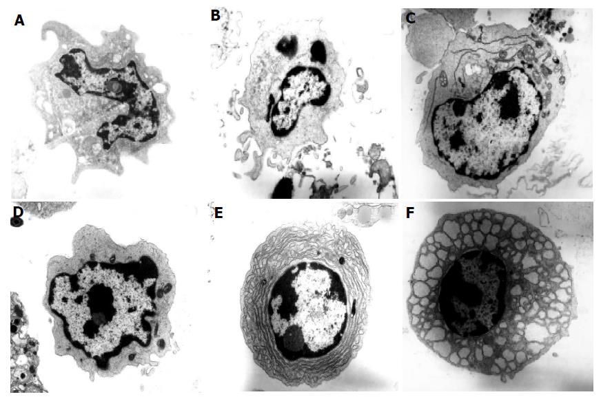
Figure 4 Electron micrographs of colonic DC from normal and colitic mice.
Electron micrographs of cells from the colonic LP of normal C57BL/6 and colitic C57BL/6-IL2-/- mice showing (A) dendritic cell type 1 (myeloid DC) from a C57BL/6 mouse (×17 250), (B) dendritic cell type 1-2 from C57BL/6-IL2-/- mouse (×17 250), (C) dendritic cell type 2 (plasmacytoid DC) from C57BL/6-IL2-/- mouse (×13 800) with prominent RER, (D) dendritic cell type 1 (myeloid DC) from C57BL/6-IL2-/- mouse (×23 000), (E) plasma cell from C57BL/6-IL2-/- mouse (×17 250) and (F) Mott cell from C57BL/6-IL2-/- mouse (×17 250). Tissue was analyzed from the colons of 3 C57BL/6-IL2-/- and 3 C57BL/6 animals.
- Citation: Cruickshank SM, English NR, Felsburg PJ, Carding SR. Characterization of colonic dendritic cells in normal and colitic mice. World J Gastroenterol 2005; 11(40): 6338-6347
- URL: https://www.wjgnet.com/1007-9327/full/v11/i40/6338.htm
- DOI: https://dx.doi.org/10.3748/wjg.v11.i40.6338


