Copyright
©2005 Baishideng Publishing Group Inc.
World J Gastroenterol. Oct 28, 2005; 11(40): 6312-6321
Published online Oct 28, 2005. doi: 10.3748/wjg.v11.i40.6312
Published online Oct 28, 2005. doi: 10.3748/wjg.v11.i40.6312
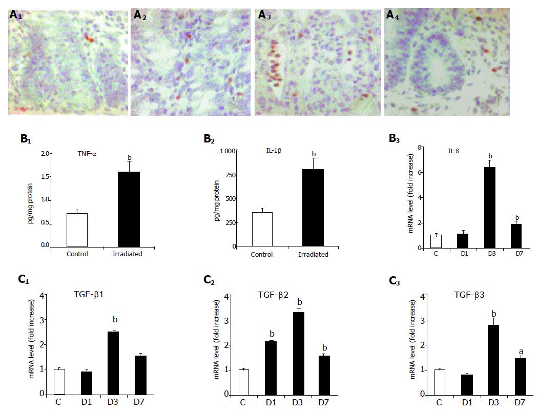
Figure 5 Radiation-induced inflammation.
A: Myeloperoxidase positive cells in X-irradiated ilea. Immunostaining for myeloperoxidase positive cells in control ilea (A1) and one (A2), three (A3) and seven (A4) days after X-irradiation respectively. Magnification x 400; B1-3: Concentration of TNF-α and IL-1β six hours, and IL-8 mRNA level one to seven days, after X-irradiation with a single dose of 10 Gy. Results are mean±SE, significantly different from controls: aP<0.05, bP<0.01 vs others; C1-3: TGF-β1, β2 and β3 mRNA level after X-irradiation with a single dose of 10 Gy. Gene expression of TGF-β1, TGF-β2 and TGF-β3, was determined by real-time RT-PCR in control ilea (C) one (D1), three (D3) and seven (D7) days after X-irradiation respectively. Results are mean±SE, significantly different from controls: aP<0.05, bP<0.01 vs others.
- Citation: Strup-Perrot C, Vozenin-Brotons MC, Vandamme M, Linard C, Mathé D. Expression of matrix metalloproteinases and tissue inhibitor metalloproteinases increases in X-irradiated rat ileum despite the disappearance of CD8a T cells. World J Gastroenterol 2005; 11(40): 6312-6321
- URL: https://www.wjgnet.com/1007-9327/full/v11/i40/6312.htm
- DOI: https://dx.doi.org/10.3748/wjg.v11.i40.6312


