Copyright
©2005 Baishideng Publishing Group Inc.
World J Gastroenterol. Oct 28, 2005; 11(40): 6312-6321
Published online Oct 28, 2005. doi: 10.3748/wjg.v11.i40.6312
Published online Oct 28, 2005. doi: 10.3748/wjg.v11.i40.6312
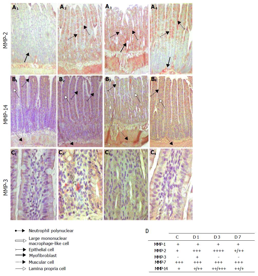
Figure 3 Tissue localization of gelatinase A and other MMPs in ilea after X-irradiation with a single dose of 10 Gy.
A2: MMP-2, MMP-14 and MMP-3 immunostaining. In control ilea, MMP-2 staining was observed in the pericryptal myofibroblast sheath, inflammatory cells and smooth muscle cells (A1, x100). On day one and three (A2; A3, x100), increased MMP-2 staining was found in all of the layer of the bowel particularly in smooth muscle cells and in epithelial cells. On day seven (A4, x100) MMP-2 staining was weaker and found in smooth muscle cells, epithelial cells of the villus only and pericryptal myofibroblast sheath. In control ilea, MMP-14 staining was observed in the inflammatory cells of the lamina propria, smooth muscle cells and some epithelial cells of the top of the villi (B1, x100). No significant modification in MMP-14 staining was found in irradiated ilea on day one (B2; x100), whereas on day three MMP-14 staining spread to the whole tissue (B3, x100). On day seven (B4, x100), MMP-14 staining return to control level except in the epithelial cells of the top of the villus. In control ilea, no MMP-3 staining was observed (C1, x400), but a staining was observed in mononuclear infiltrated cells day one after X-irradiation (C2, x400); D: MMPs immunostaining scoring. (-) represent no staining, (+) weak staining, (++) moderate staining, (+++) important and (++++) strong staining.
- Citation: Strup-Perrot C, Vozenin-Brotons MC, Vandamme M, Linard C, Mathé D. Expression of matrix metalloproteinases and tissue inhibitor metalloproteinases increases in X-irradiated rat ileum despite the disappearance of CD8a T cells. World J Gastroenterol 2005; 11(40): 6312-6321
- URL: https://www.wjgnet.com/1007-9327/full/v11/i40/6312.htm
- DOI: https://dx.doi.org/10.3748/wjg.v11.i40.6312


