Copyright
©2005 Baishideng Publishing Group Inc.
World J Gastroenterol. Jan 28, 2005; 11(4): 609-611
Published online Jan 28, 2005. doi: 10.3748/wjg.v11.i4.609
Published online Jan 28, 2005. doi: 10.3748/wjg.v11.i4.609
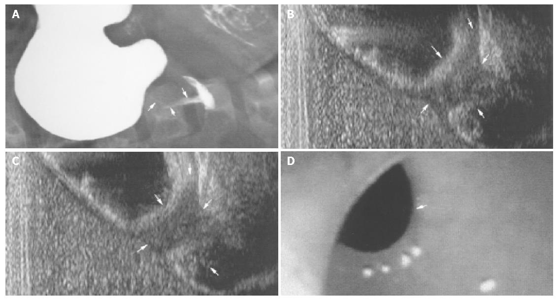
Figure 1 Clinical findings in antral web associated with antral hyperatrophy and prepyloric stenosis.
A: Barium meal study showing a poorly expanded distal antral lumen (arrows) and pyloric canal (open arrow) mimicking hypertrophic pyloric stenosis; B: Abdominal ultrasound of the stomach after water loading showing an echogenic antral flap (large open arrow) with an eccentric aperture (white triangle), thickened antral wall (arrows) beyond the flap, and lack of flow (small open arrow) through the antropyloric regionl; C: Abdominal ultrasound of the stomach with compression on the upper abdomen showing the antral flap (large open arrow) and thickened distal antral wall (arrows), turbulent flow at the aperture (black triangle), and jet-like flow (small open arrow) through the antropyloric region to the duodenal bulb; D: Endoscopy of the stomach confirming the presence of an antral web with an eccentric aperture (arrow).
- Citation: Tiao MM, Ko SF, Hsieh CS, Ng SH, Liang CD, Sheen-Chen SM, Chuang JH, Huang HY. Antral web associated with distal antral hypertrophy and prepyloric stenosis mimicking hypertrophic pyloric stenosis. World J Gastroenterol 2005; 11(4): 609-611
- URL: https://www.wjgnet.com/1007-9327/full/v11/i4/609.htm
- DOI: https://dx.doi.org/10.3748/wjg.v11.i4.609


