Copyright
©2005 Baishideng Publishing Group Inc.
World J Gastroenterol. Sep 21, 2005; 11(35): 5485-5491
Published online Sep 21, 2005. doi: 10.3748/wjg.v11.i35.5485
Published online Sep 21, 2005. doi: 10.3748/wjg.v11.i35.5485
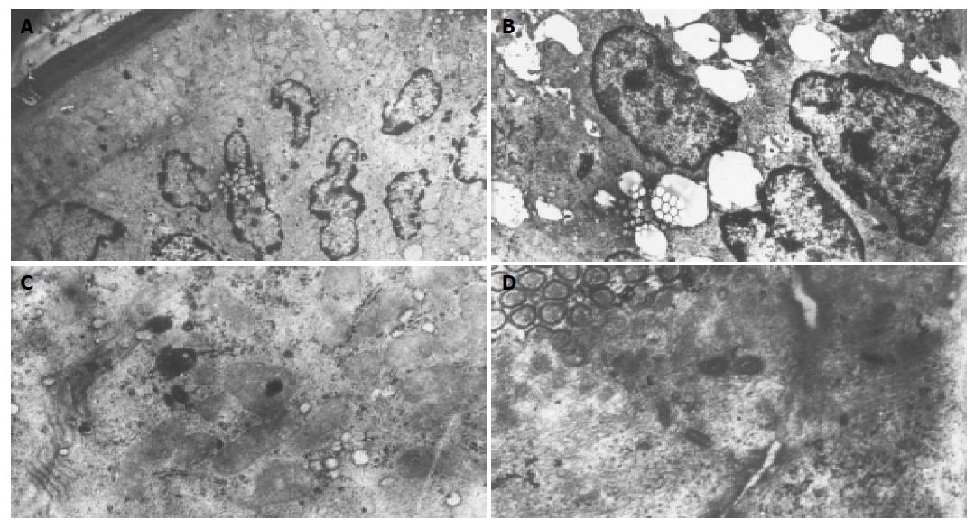
Figure 2 Electron microscopic examination of ileum following ischemia-reperfusion injury after resuscitation of hemorrhagic shock.
The apoptosis and necrosis were main manifestation in the intestinal mucosal injury. A: Many enterocytes show compaction and segregation of chromatin against the nuclear envelope, but still preserve mcrovilli and intact mitochondria; B: Showing the appearance of membrane blebs which cause detachment from the basement membrane; C: Some enterocytes show the swelling of mitochondria and endoplasmic reticulum, scarce and lodging microvilli; D: the opening of tight junction among the cells. Original magnification: ×2 500 (A, B), ×4 000 (C,D).
- Citation: Chang JX, Chen S, Ma LP, Jiang LY, Chen JW, Chang RM, Wen LQ, Wu W, Jiang ZP, Huang ZT. Functional and morphological changes of the gut barrier during the restitution process after hemorrhagic shock. World J Gastroenterol 2005; 11(35): 5485-5491
- URL: https://www.wjgnet.com/1007-9327/full/v11/i35/5485.htm
- DOI: https://dx.doi.org/10.3748/wjg.v11.i35.5485


