Copyright
©2005 Baishideng Publishing Group Inc.
World J Gastroenterol. Sep 21, 2005; 11(35): 5468-5474
Published online Sep 21, 2005. doi: 10.3748/wjg.v11.i35.5468
Published online Sep 21, 2005. doi: 10.3748/wjg.v11.i35.5468
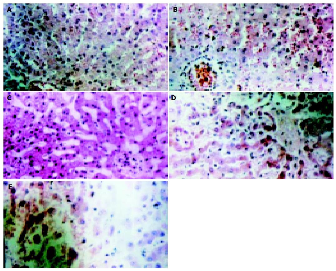
Figure 2 Liver histology observation at 0 h (A), 1 h (B), 4 h (C), 7 h (D) after liver devascularization, and death of pigs (E) under light microscope.
- Citation: Gao Y, Mu N, Xu XP, Wang Y. Porcine acute liver failure model established by two-phase surgery and treated with hollow fiber bioartificial liver support system. World J Gastroenterol 2005; 11(35): 5468-5474
- URL: https://www.wjgnet.com/1007-9327/full/v11/i35/5468.htm
- DOI: https://dx.doi.org/10.3748/wjg.v11.i35.5468


