Copyright
©The Author(s) 2005.
World J Gastroenterol. Sep 14, 2005; 11(34): 5416-5417
Published online Sep 14, 2005. doi: 10.3748/wjg.v11.i34.5416
Published online Sep 14, 2005. doi: 10.3748/wjg.v11.i34.5416
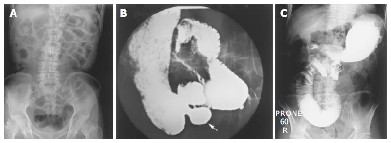
Figure 1 A plain X-ray of the abdomen showing dilatation of the small intestine, especially the proximal jejunum (A).
B and C: An upper GI oral contrast study showing multiple diverticula in the duodenum and proximal jejunum (the white arrow, (B) and dilatation of the proximal jejunum (C).
- Citation: Lin CH, Hsieh HF, Yu CY, Yu JC, Chan DC, Chen TW, Chen PJ, Liu YC. Diverticulosis of the jejunum with intestinal obstruction: A case report. World J Gastroenterol 2005; 11(34): 5416-5417
- URL: https://www.wjgnet.com/1007-9327/full/v11/i34/5416.htm
- DOI: https://dx.doi.org/10.3748/wjg.v11.i34.5416


