Copyright
©The Author(s) 2005.
World J Gastroenterol. Sep 14, 2005; 11(34): 5336-5341
Published online Sep 14, 2005. doi: 10.3748/wjg.v11.i34.5336
Published online Sep 14, 2005. doi: 10.3748/wjg.v11.i34.5336
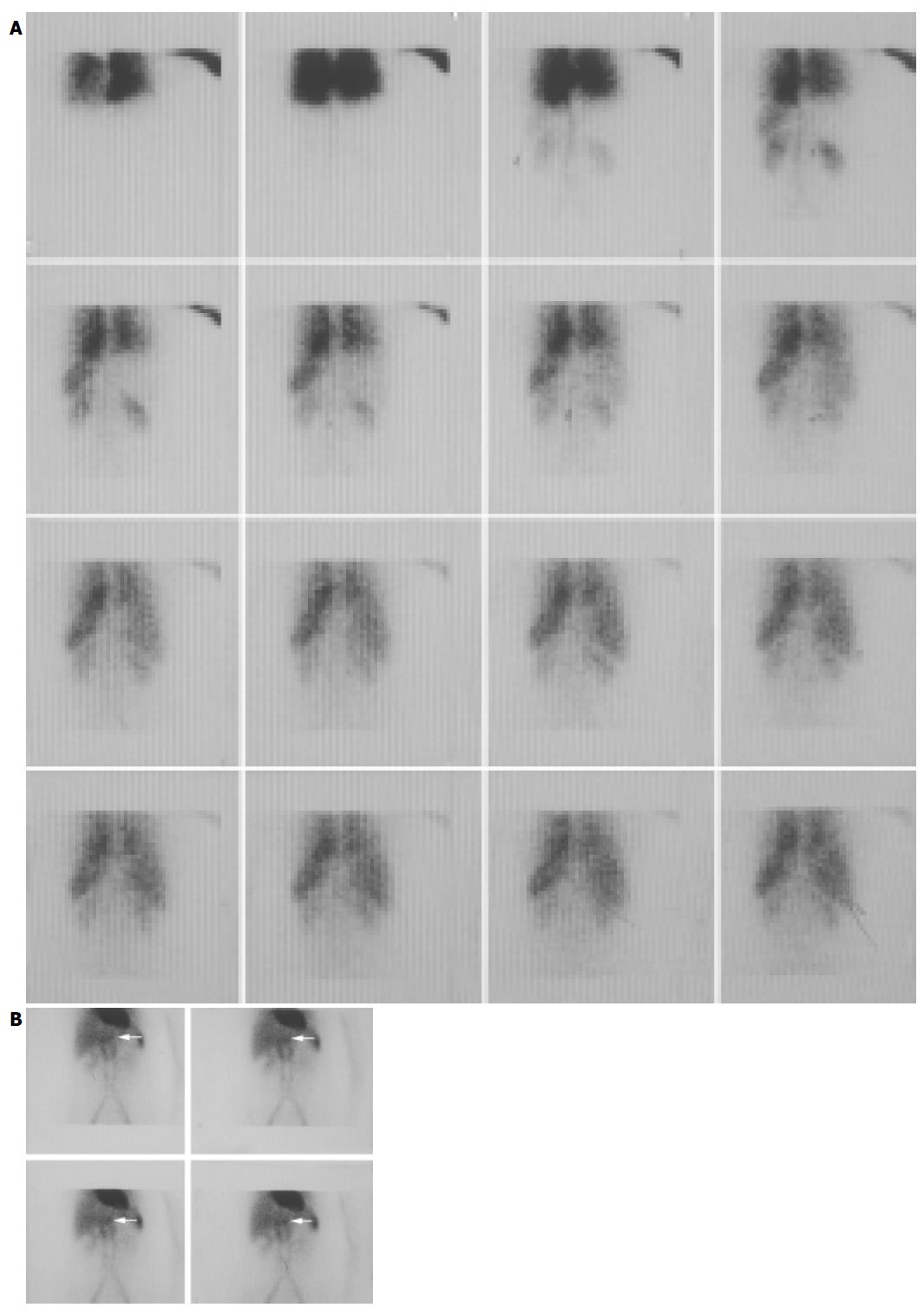
Figure 1 Female, 46 years old.
US showed that there was a lesion in the left liver with the size 3.6×2.3 cm2. The function of liver was normal. SPECT/CT test was carried out to exclude hepatic hemangioma. A: The hepatic blood perfusion imaging. The abnormal focal accumulation of the radioactivity was not shown obviously; B: The radioactivity accumulation of the focus (pointed by the red arrows) in the left liver was a little higher than the normal liver tissue.
- Citation: Zheng JG, Yao ZM, Shu CY, Zhang Y, Zhang X. Role of SPECT/CT in diagnosis of hepatic hemangiomas. World J Gastroenterol 2005; 11(34): 5336-5341
- URL: https://www.wjgnet.com/1007-9327/full/v11/i34/5336.htm
- DOI: https://dx.doi.org/10.3748/wjg.v11.i34.5336


