Copyright
©The Author(s) 2005.
World J Gastroenterol. Sep 7, 2005; 11(33): 5203-5208
Published online Sep 7, 2005. doi: 10.3748/wjg.v11.i33.5203
Published online Sep 7, 2005. doi: 10.3748/wjg.v11.i33.5203
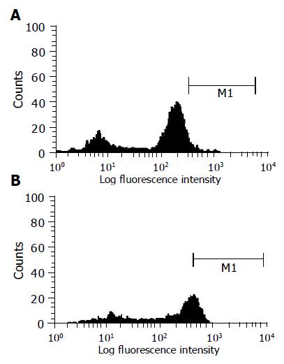
Figure 2 Histogram from gated leukocytes obtained from fluorescent activated cell sorter analysis of HCV-infected peripheral blood mononuclear leukocytes.
Cells were stained intracellularly with anti-C2 antibody conjugated with FITC. Histogram represents gated leukocytes from (A) healthy uninfected cells or (B) infected cells stained with anti-C2 antibody conjugated with FITC after incubation of blood with serum sample after 24 h at 37 °C in which x-axis represents fluorescence intensity. M1 is marker for positive cell population.
- Citation: El-Awady MK, Tabll AA, Redwan ERM, Youssef S, Omran MH, Thakeb F, El-Demellawy M. Flow cytometric detection of hepatitis C virus antigens in infected peripheral blood leukocytes: Binding and entry. World J Gastroenterol 2005; 11(33): 5203-5208
- URL: https://www.wjgnet.com/1007-9327/full/v11/i33/5203.htm
- DOI: https://dx.doi.org/10.3748/wjg.v11.i33.5203


