Copyright
©The Author(s) 2005.
World J Gastroenterol. Sep 7, 2005; 11(33): 5095-5102
Published online Sep 7, 2005. doi: 10.3748/wjg.v11.i33.5095
Published online Sep 7, 2005. doi: 10.3748/wjg.v11.i33.5095
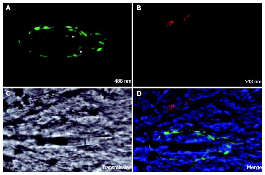
Figure 2 CC531s were labeled with DiO, injected into the mesenteric vein.
After 24 h, Kupffer cells (KC) were stained in vivo with TRITC-labeled latex beads, and the liver was fixed by perfusion. Confocal microscopy was conducted and images were named after corresponding laser lines. A: Group of CC531s was visualized; B: KC was visualized by the up-take of fluorescent latex beads; C: The rest of the field was examined with the transmission function; D: CC531s proliferating inside the liver parenchyma and inside the group of CC531s cells, a vessel was seen. The vessel is much straighter than the normal liver sinusoids, indicating a newly formed vessel. Image size: 158.7 μm×158.7 μm.
- Citation: Vekemans K, Braet F. Structural and functional aspects of the liver and liver sinusoidal cells in relation to colon carcinoma metastasis. World J Gastroenterol 2005; 11(33): 5095-5102
- URL: https://www.wjgnet.com/1007-9327/full/v11/i33/5095.htm
- DOI: https://dx.doi.org/10.3748/wjg.v11.i33.5095


