Copyright
©The Author(s) 2005.
World J Gastroenterol. Sep 7, 2005; 11(33): 5095-5102
Published online Sep 7, 2005. doi: 10.3748/wjg.v11.i33.5095
Published online Sep 7, 2005. doi: 10.3748/wjg.v11.i33.5095
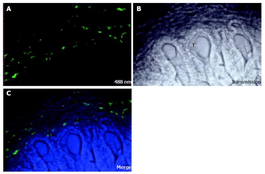
Figure 1 CC531s cells were injected into the mesenteric vein and were left in circulation for 17 d.
One hour before perfusion fixation, FSA was injected into the penile vein. FSA stains only LSECs. Confocal microscopy was conducted and images were named after corresponding laser lines. A: FSA staining was measured with CLSM; fluorescence was observed only in part of the scanned field; B: Transmission image of the same field, with the tumor region in the lower part of the field. Formation of cryptae of the tumor (T) can be observed; C: After merging the two images, no staining was observed inside the tumor region. Image size: 500 μm×500 μm.
- Citation: Vekemans K, Braet F. Structural and functional aspects of the liver and liver sinusoidal cells in relation to colon carcinoma metastasis. World J Gastroenterol 2005; 11(33): 5095-5102
- URL: https://www.wjgnet.com/1007-9327/full/v11/i33/5095.htm
- DOI: https://dx.doi.org/10.3748/wjg.v11.i33.5095


