Copyright
©The Author(s) 2005.
World J Gastroenterol. Aug 28, 2005; 11(32): 5079-5081
Published online Aug 28, 2005. doi: 10.3748/wjg.v11.i32.5079
Published online Aug 28, 2005. doi: 10.3748/wjg.v11.i32.5079
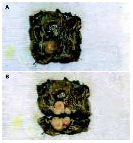
Figure 1 Macroscopic view of the sigmoid colon showed a well-circumscribed submucosal tumor (A).
The cut surface of this tumor consisted of fibrotic and myxoid parts and was gray-yellowish in color (B).
- Citation: Fotiadis CI, Kouerinis IA, Papandreou I, Zografos GC, Agapitos G. Sigmoid schwannoma: A rare case. World J Gastroenterol 2005; 11(32): 5079-5081
- URL: https://www.wjgnet.com/1007-9327/full/v11/i32/5079.htm
- DOI: https://dx.doi.org/10.3748/wjg.v11.i32.5079


