Copyright
©The Author(s) 2005.
World J Gastroenterol. Aug 28, 2005; 11(32): 4939-4946
Published online Aug 28, 2005. doi: 10.3748/wjg.v11.i32.4939
Published online Aug 28, 2005. doi: 10.3748/wjg.v11.i32.4939
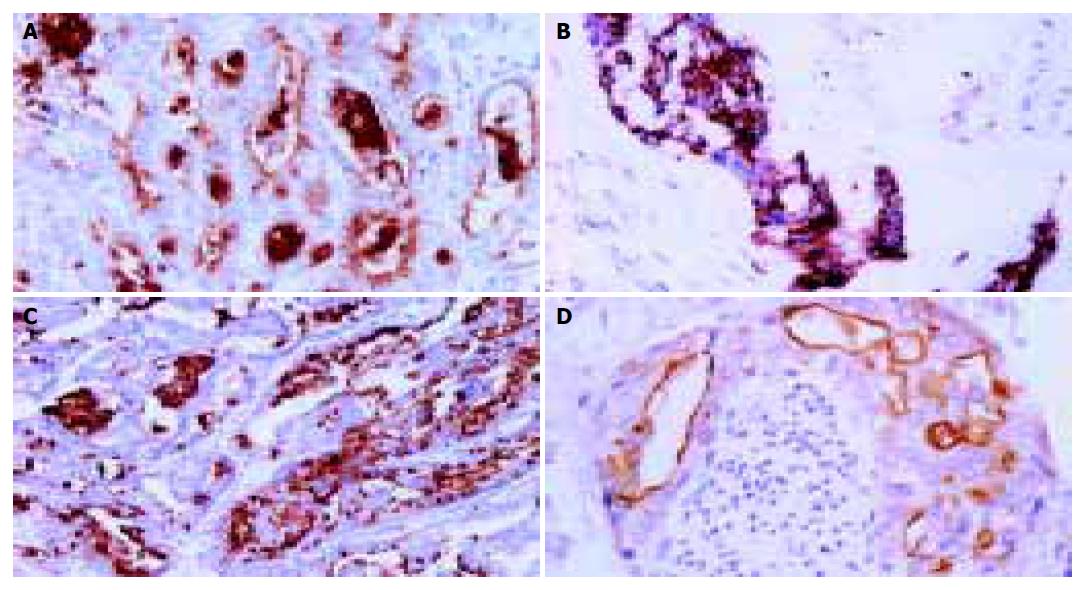
Figure 1 Staining patterns of MUC1 and MUC5AC core proteins.
A and C: Immunohistochemical staining of MUC1 and MUC5AC apomucins expressed in tumor tissues, respectively. B and D: MUC1 and MUC5AC staining of vascular and neural-invaded tumor cells.
- Citation: Boonla C, Sripa B, Thuwajit P, Cha-On U, Puapairoj A, Miwa M, Wongkham S. MUC1 and MUC5AC mucin expression in liver fluke-associated intrahepatic cholangiocarcinoma. World J Gastroenterol 2005; 11(32): 4939-4946
- URL: https://www.wjgnet.com/1007-9327/full/v11/i32/4939.htm
- DOI: https://dx.doi.org/10.3748/wjg.v11.i32.4939


