Copyright
©The Author(s) 2005.
World J Gastroenterol. Aug 14, 2005; 11(30): 4611-4617
Published online Aug 14, 2005. doi: 10.3748/wjg.v11.i30.4611
Published online Aug 14, 2005. doi: 10.3748/wjg.v11.i30.4611
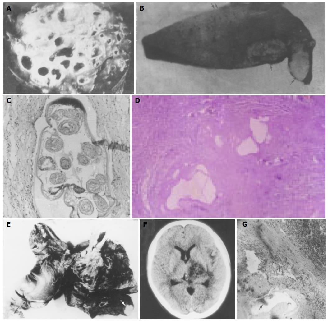
Figure 7 Pathology.
A: A cross section of human liver AE showed alveolate appearance; B: Types of liver AE.↑: Nodular type, ↑↑: Mixed type; C: Protoscolices inside brood capsule (HE stain×100); D: Alveococcus nodule (HE stain×200); E: Three arrows indicating metastatic bilateral lung AE; F: ↑: metastatic right brain AE; G: lymph node metastasis near porta hepatis (HE stain×200). ↑: AE. The left upper side: Metastatic lymph nodes.
- Citation: Jiang CP, Don M, Jones M. Liver alveolar echinococcosis in China: Clinical aspect with relative basic research. World J Gastroenterol 2005; 11(30): 4611-4617
- URL: https://www.wjgnet.com/1007-9327/full/v11/i30/4611.htm
- DOI: https://dx.doi.org/10.3748/wjg.v11.i30.4611


