Copyright
©The Author(s) 2005.
World J Gastroenterol. Aug 14, 2005; 11(30): 4611-4617
Published online Aug 14, 2005. doi: 10.3748/wjg.v11.i30.4611
Published online Aug 14, 2005. doi: 10.3748/wjg.v11.i30.4611
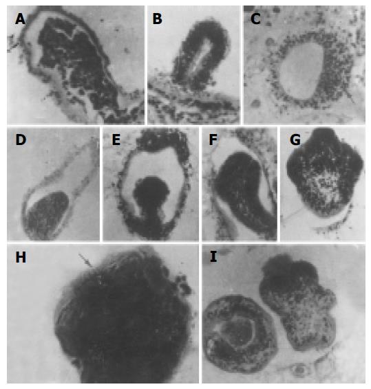
Figure 5 The entire process of mouse AE protoscolex histogenesis (HE stain×200-800).
A: local cellular hyperplasia of germinal membrane of alveococcus wall; B: re-arrangement of hyperplastic cells and formation of brood capsule; C: local cellular hyperplasia of brood capsule wall; D: elliptical protrusion into cavity; E: mushroom protrusion; F: lingural protrusion; G: showing rostellum and suckers of protoscolex; H: Hooklets on rostellum; I: mature protoscolex (right: evaginated type. Left: invaginated type).
- Citation: Jiang CP, Don M, Jones M. Liver alveolar echinococcosis in China: Clinical aspect with relative basic research. World J Gastroenterol 2005; 11(30): 4611-4617
- URL: https://www.wjgnet.com/1007-9327/full/v11/i30/4611.htm
- DOI: https://dx.doi.org/10.3748/wjg.v11.i30.4611


