Copyright
©The Author(s) 2005.
World J Gastroenterol. Aug 7, 2005; 11(29): 4607-4609
Published online Aug 7, 2005. doi: 10.3748/wjg.v11.i29.4607
Published online Aug 7, 2005. doi: 10.3748/wjg.v11.i29.4607
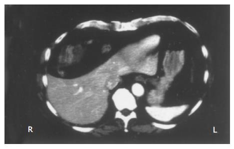
Figure 1 Axial CT scan of the abdomen.
The image shows interposition of the colon (with fecal content) between the rib cage and the liver (Chilaiditi’s sign). The latter appears compressed and dislocated in the absence of parenchymal and focal lesions.
- Citation: Sorrentino D, Bazzocchi M, Badano L, Toso F, Giagu P. Heart-touching Chilaiditi’s syndrome. World J Gastroenterol 2005; 11(29): 4607-4609
- URL: https://www.wjgnet.com/1007-9327/full/v11/i29/4607.htm
- DOI: https://dx.doi.org/10.3748/wjg.v11.i29.4607


