Copyright
©The Author(s) 2005.
World J Gastroenterol. Aug 7, 2005; 11(29): 4505-4510
Published online Aug 7, 2005. doi: 10.3748/wjg.v11.i29.4505
Published online Aug 7, 2005. doi: 10.3748/wjg.v11.i29.4505
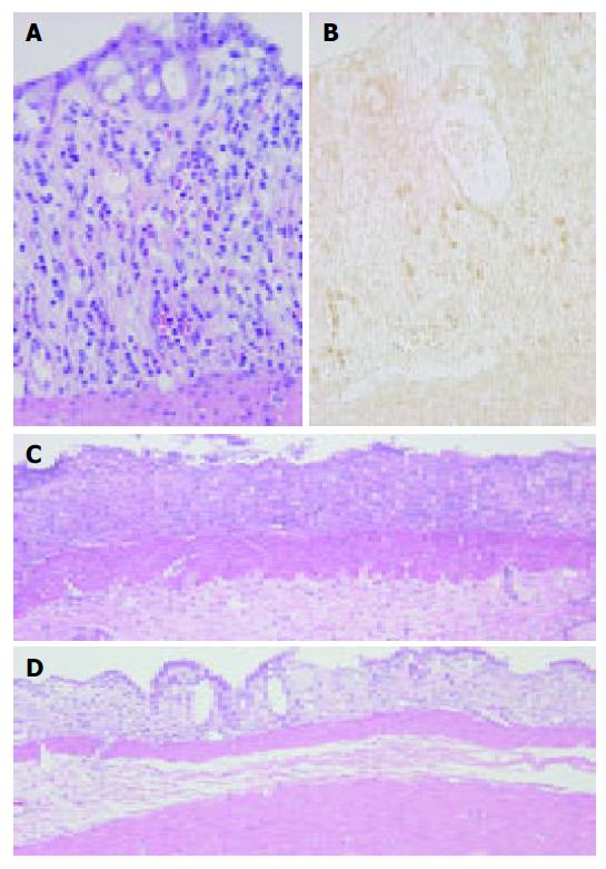
Figure 2 Colonic mucosa at 3 d post-DSS treatment showing eosinophils in the lamina propria.
HE staining (A) and immunostaining for ECP (B) as described under Materials and methods. ECP-positive cells (brown) appear in close proximity to damaged crypts in the lamina propria and partially in the extracellular interstitium of DSS-induced colitis. Original magnification, ×400. Colonic mucosa at 7-d treatment of DSS. (C) Colonic ulceration in normal serum-treated rats. (D) Reduced severity of colonic mucosal ulceration in anti-ECP-treated rats. HE stain. Original magnification, ×100.
- Citation: Shichijo K, Makiyama K, Wen CY, Matsuu M, Nakayama T, Nakashima M, Ihara M, Sekine I. Antibody to eosinophil cationic protein suppresses dextran sulfate sodium-induced colitis in rats. World J Gastroenterol 2005; 11(29): 4505-4510
- URL: https://www.wjgnet.com/1007-9327/full/v11/i29/4505.htm
- DOI: https://dx.doi.org/10.3748/wjg.v11.i29.4505


