Copyright
©The Author(s) 2005.
World J Gastroenterol. Aug 7, 2005; 11(29): 4497-4504
Published online Aug 7, 2005. doi: 10.3748/wjg.v11.i29.4497
Published online Aug 7, 2005. doi: 10.3748/wjg.v11.i29.4497
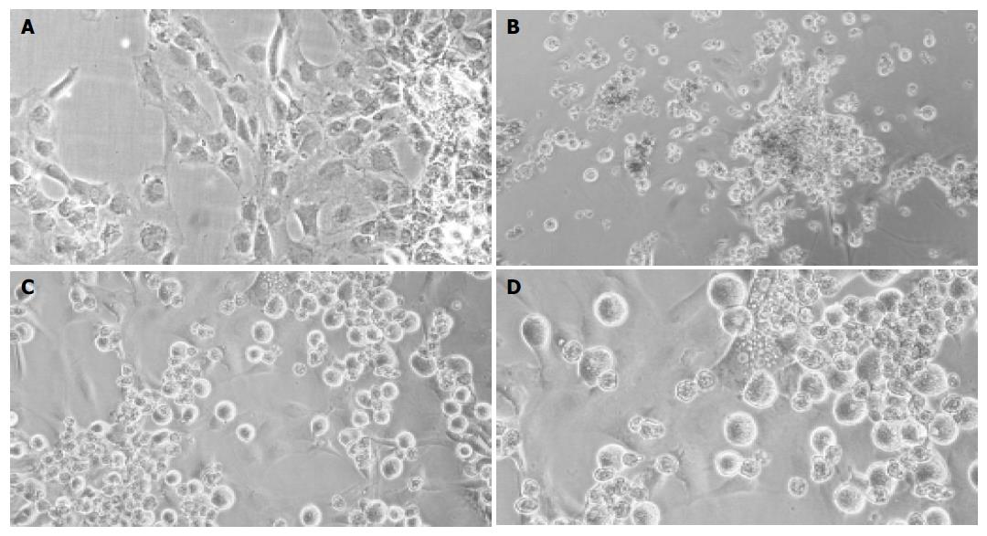
Figure 2 Light-microscopic pictures of cultured cells.
Cultured rMSC (A), hepatocyte controls (B), or cocultures of rMSC with hepatocytes (C + enlargement D) after 3 wk. Cultured rMSC grew adherent and adopted a polygonal cell morphology after weeks in culture (A). Hepatocytes formed clumps of rounded cells, and few viable cells were observed after 3 wk in culture (B). In mixed cultures, mainly MSC attached to the substratum, whereas hepatocytes grew over the MSC-layer forming clusters (C). Binucleated cells were found within the attached layer of the cultured MSC beginning with 1 wk in culture. Shown is one example at wk 2 out of three experiments. Original magnification ×200.
- Citation: Lange C, Bassler P, Lioznov MV, Bruns H, Kluth D, Zander AR, Fiegel HC. Liver-specific gene expression in mesenchymal stem cells is induced by liver cells. World J Gastroenterol 2005; 11(29): 4497-4504
- URL: https://www.wjgnet.com/1007-9327/full/v11/i29/4497.htm
- DOI: https://dx.doi.org/10.3748/wjg.v11.i29.4497


