Copyright
©The Author(s) 2005.
World J Gastroenterol. Aug 7, 2005; 11(29): 4490-4496
Published online Aug 7, 2005. doi: 10.3748/wjg.v11.i29.4490
Published online Aug 7, 2005. doi: 10.3748/wjg.v11.i29.4490
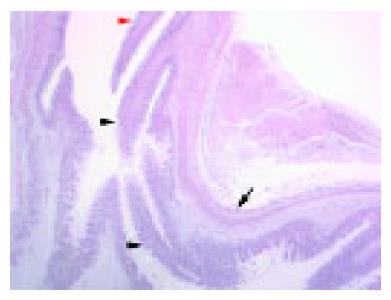
Figure 2 Overview of the fetal EGJ at 20-wk GA.
The upper border of the abdominal esophagus is indicated by a red arrowhead. The angle of His (the anatomic boundary between the tubular esophagus and the saccular stomach) is easily recognizable (arrow). The distal end of the esophagus is located at the level of the angle of His. Esophageal simple columnar epithelium is present in the distal esophagus (indicated by black arrowheads at its proximal and distal margins, H&E, OM ×125).
- Citation: Hertogh GD, Eyken PV, Ectors N, Geboes K. On the origin of cardiac mucosa: A histological and immunohistoc-hemical study of cytokeratin expression patterns in the developing esophagogastric junction region and stomach. World J Gastroenterol 2005; 11(29): 4490-4496
- URL: https://www.wjgnet.com/1007-9327/full/v11/i29/4490.htm
- DOI: https://dx.doi.org/10.3748/wjg.v11.i29.4490


