Copyright
©The Author(s) 2005.
World J Gastroenterol. Jul 28, 2005; 11(28): 4382-4389
Published online Jul 28, 2005. doi: 10.3748/wjg.v11.i28.4382
Published online Jul 28, 2005. doi: 10.3748/wjg.v11.i28.4382
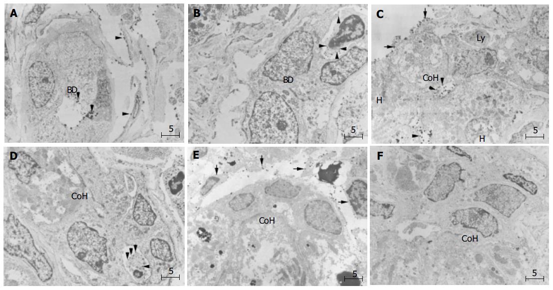
Figure 5 (PDF) Immunoelectron microscopic findings of ICAM-1 ( A, C, and E) and LFA-1 ( B, D, and F) in periportal small bile ductule and canal of Hering in PBC liver.
A: Gold-labeled ICAM-1 particles on the surface of cholangiocytes (arrowhead) facing the lumen of small bile ductule, a small portal or peripheral blood vessel, and also on sinusoidal endothelial cells (arrowheads). BD denotes bile ductule; B: Lymphocytes with dense labeling of LFA-1 (arrowhead) around small ductule; C: Gold-labeled ICAM-1 particles on the luminal surfaces of hepatocytes and cholangiocytes of CoH, on bile canaliculus and also on sinusoidal endothelial cells (arrows); D: Lymphocytes densely labeled with LFA-1 (arrows) on the basolateral membrane of cholangiocytes of the CoH; E:No ICAM-1 immunoreactivity show immature cells resembling progenitor cells (asterisk) in CoH, and these cells; F: No infiltration of lymphocytes is observed around the progenitor-like cells (asterisk). Bar, 5 mm; BD, bile ductile; Ly, lymphocyte; CoH, canal of Hering; H. hepatocyte. Uranyl acetate stain.
- Citation: Yokomori H, Oda M, Ogi M, Wakabayashi G, Kawachi S, Yoshimura K, Nagai T, Kitajima M, Nomura M, Hibi T. Expression of adhesion molecules on mature cholangiocytes in canal of Hering and bile ductules in wedge biopsy samples of primary biliary cirrhosis. World J Gastroenterol 2005; 11(28): 4382-4389
- URL: https://www.wjgnet.com/1007-9327/full/v11/i28/4382.htm
- DOI: https://dx.doi.org/10.3748/wjg.v11.i28.4382


