Copyright
©The Author(s) 2005.
World J Gastroenterol. Jul 21, 2005; 11(27): 4258-4260
Published online Jul 21, 2005. doi: 10.3748/wjg.v11.i27.4258
Published online Jul 21, 2005. doi: 10.3748/wjg.v11.i27.4258
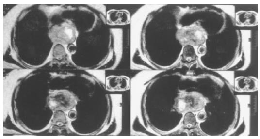
Figure 1 A 42-year-old male with giant esophageal leiomyoma.
MRI image shows the tumor located in lower thirds of the esophagus. The size of tumor is 10 cm × 10 cm × 6 cm.
- Citation: Cheng BC, Chang S, Mao ZF, Li MJ, Huang J, Wang ZW, Wang TS. Surgical treatment of giant esophageal leiomyoma. World J Gastroenterol 2005; 11(27): 4258-4260
- URL: https://www.wjgnet.com/1007-9327/full/v11/i27/4258.htm
- DOI: https://dx.doi.org/10.3748/wjg.v11.i27.4258


