Copyright
©The Author(s) 2005.
World J Gastroenterol. Jul 14, 2005; 11(26): 4120-4123
Published online Jul 14, 2005. doi: 10.3748/wjg.v11.i26.4120
Published online Jul 14, 2005. doi: 10.3748/wjg.v11.i26.4120
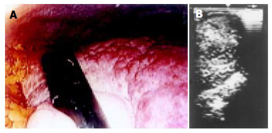
Figure 2 A: Endoscopic liver image of cirrhosis.
Ultrasonic probe is placed in contact to a fibrotic depressed whitish area near the gallbladder; B: Laparoscopic sonogram revealing a tumor under liver surface.
- Citation: Gómez-Rubio M, Moya-Valdés M, García J. Diagnostic laparoscopy and laparoscopic ultrasonography with local anesthesia in hepatocellular carcinoma. World J Gastroenterol 2005; 11(26): 4120-4123
- URL: https://www.wjgnet.com/1007-9327/full/v11/i26/4120.htm
- DOI: https://dx.doi.org/10.3748/wjg.v11.i26.4120


