Copyright
©The Author(s) 2005.
World J Gastroenterol. Jul 14, 2005; 11(26): 4117-4119
Published online Jul 14, 2005. doi: 10.3748/wjg.v11.i26.4117
Published online Jul 14, 2005. doi: 10.3748/wjg.v11.i26.4117
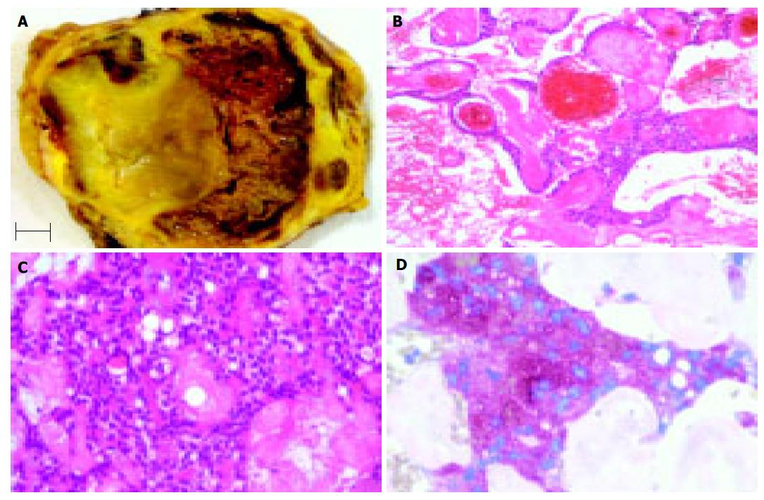
Figure 1 Cross section of the pancreatic tumor showed a yellow tumor with hemorrhages (A); bar indicates 1 cm.
The tumor tissue exhibits a solid monomorphic pattern with a pseudopapillary pattern in areas with regressive changes (B) and small polyhedral cells lining fibrovascular stalks (C). The tumor cells were immunoreactive for alpha-1-antitrypsin (D). Hematoxylin and eosin (B and C), anti-alpha-1-antitrypsin (D); Original magnification: x100 (B), x200 (C), x400 (D).
- Citation: Eder F, Schulz HU, Röcken C, Lippert H. Solid-pseudopapillary tumor of the pancreatic tail. World J Gastroenterol 2005; 11(26): 4117-4119
- URL: https://www.wjgnet.com/1007-9327/full/v11/i26/4117.htm
- DOI: https://dx.doi.org/10.3748/wjg.v11.i26.4117


