Copyright
©The Author(s) 2005.
World J Gastroenterol. Jul 14, 2005; 11(26): 4024-4031
Published online Jul 14, 2005. doi: 10.3748/wjg.v11.i26.4024
Published online Jul 14, 2005. doi: 10.3748/wjg.v11.i26.4024
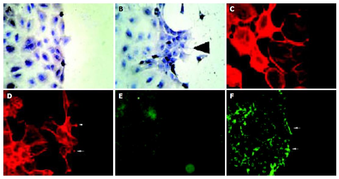
Figure 6 Stimulation of IEC-6 cells with EphB1-Fc induces subcellular responses compatible with enhanced wound closure capacity.
Compared to the non-stimulated control (A), recombinant EphB1-Fc stimulated cells produce an organized migratory activity forming multiple archipelago-like sheets of cells stretching into the denuded area already 6 h after stimulation. In addition, asymmetric lamellipodia are seen (arrow). (B). Staining of actin with phalloidin shows a rounded morphology with less polymerized actin bundles in unstimulated cells (C), whereas EphB1-Fc stimulated cells display a stretched-out, bizarre morphology and apparent assembly of abundant stress fibers. Spike-like protrusions may correspond to focal adhesion assembly (arrows) (D). EphB1-Fc also induces an increased deposition of laminin as a provisional basal membrane at the wound edge (F) (arrows) compared to control cells (E).
- Citation: Hafner C, Meyer S, Langmann T, Schmitz G, Bataille F, Hagen I, Becker B, Roesch A, Rogler G, Landthaler M, Vogt T. Ephrin-B2 is differentially expressed in the intestinal epithelium in Crohn’s disease and contributes to accelerated epithelial wound healing in vitro. World J Gastroenterol 2005; 11(26): 4024-4031
- URL: https://www.wjgnet.com/1007-9327/full/v11/i26/4024.htm
- DOI: https://dx.doi.org/10.3748/wjg.v11.i26.4024


