Copyright
©2005 Baishideng Publishing Group Inc.
World J Gastroenterol. Jun 28, 2005; 11(24): 3791-3793
Published online Jun 28, 2005. doi: 10.3748/wjg.v11.i24.3791
Published online Jun 28, 2005. doi: 10.3748/wjg.v11.i24.3791
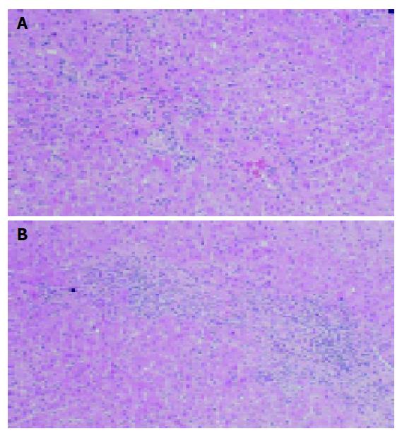
Figure 2 Liver biopsy.
A: Focal necrosis of the parenchyma was observed; B: Portal spaces were slightly enlarged with mild fibrosis and were occupied with infiltrates including a few plasma cells. The limiting plate was mostly preserved. Hematoxylin-eosin staining; original magnification ×200.
- Citation: Mikata R, Yokosuka O, Imazeki F, Fukai K, Kanda T, Saisho H. Prolonged acute hepatitis A mimicking autoimmune hepatitis. World J Gastroenterol 2005; 11(24): 3791-3793
- URL: https://www.wjgnet.com/1007-9327/full/v11/i24/3791.htm
- DOI: https://dx.doi.org/10.3748/wjg.v11.i24.3791


