Copyright
©2005 Baishideng Publishing Group Inc.
World J Gastroenterol. Jun 14, 2005; 11(22): 3368-3374
Published online Jun 14, 2005. doi: 10.3748/wjg.v11.i22.3368
Published online Jun 14, 2005. doi: 10.3748/wjg.v11.i22.3368
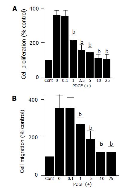
Figure 3 EGCG inhibited PDGF-induced proliferation and migration.
A: Serum-starved PSCs were left untreated (“Cont”) or treated with PDGF-BB (at 25 μg/L) in the presence or absence of EGCG at the indicated concentrations (μmol/L). After 24-h incubation, DNA synthesis was assessed by BrdU incorporation enzyme-linked immunosorbent assay. Data are shown as mean±SD (% of the control, n = 6). bP<0.01 vs PDGF only; B: cell migration was assessed using modified Boyden chambers with 8-μm pore filters. Serum-starved PSCs were left untreated (“Cont”) or were treated with PDGF-BB (at 25 μg/L) in the lower chamber in the absence or presence of EGCG at the indicated concentrations (μmol/L). After 24-h incubation with PDGF, the cells migrated to the underside of the filter were stained, and counted. Data are shown as mean±SD (% of the control, n = 6). bP<0.01 vs PDGF only.
- Citation: Masamune A, Kikuta K, Satoh M, Suzuki N, Shimosegawa T. Green tea polyphenol epigallocatechin-3-gallate blocks PDGF-induced proliferation and migration of rat pancreatic stellate cells. World J Gastroenterol 2005; 11(22): 3368-3374
- URL: https://www.wjgnet.com/1007-9327/full/v11/i22/3368.htm
- DOI: https://dx.doi.org/10.3748/wjg.v11.i22.3368


