Copyright
©2005 Baishideng Publishing Group Co.
World J Gastroenterol. Jan 14, 2005; 11(2): 249-254
Published online Jan 14, 2005. doi: 10.3748/wjg.v11.i2.249
Published online Jan 14, 2005. doi: 10.3748/wjg.v11.i2.249
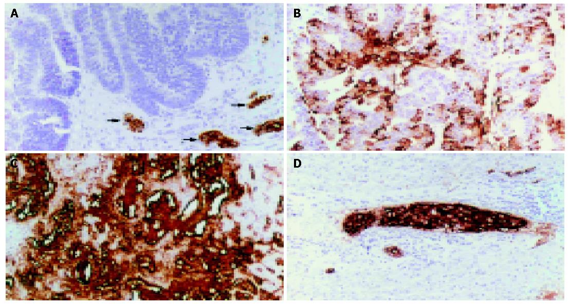
Figure 1 Immunohistochemical detection of sLea in CCA.
A: A CCA case with no sLea expression in tumor but with positive staining in the normal bile ducts (arrows); B: and C: CCA cases with positive sLea, showing apical and stromal staining, respectively; D: Vascular metastasis of sLea positive CCA cells. (Immunoperoxidase staining, original magnification ×100).
- Citation: Juntavee A, Sripa B, Pugkhem A, Khuntikeo N, Wongkham S. Expression of sialyl Lewisa relates to poor prognosis in cholangiocarcinoma. World J Gastroenterol 2005; 11(2): 249-254
- URL: https://www.wjgnet.com/1007-9327/full/v11/i2/249.htm
- DOI: https://dx.doi.org/10.3748/wjg.v11.i2.249


