Copyright
©2005 Baishideng Publishing Group Inc.
World J Gastroenterol. May 21, 2005; 11(19): 3008-3009
Published online May 21, 2005. doi: 10.3748/wjg.v11.i19.3008
Published online May 21, 2005. doi: 10.3748/wjg.v11.i19.3008
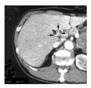
Figure 1 An abdominal CT showed compression from the dorsum and stenosis of the EBD (thick arrow head: EBD) by right hepatic artery (thin arrow head: anterior branch and posterior branch), but did not reveal a mass or lymph node swelling around the EBD.
- Citation: Miyashita K, Shiraki K, Ito T, Taoka H, Nakano T. The right hepatic artery syndrome. World J Gastroenterol 2005; 11(19): 3008-3009
- URL: https://www.wjgnet.com/1007-9327/full/v11/i19/3008.htm
- DOI: https://dx.doi.org/10.3748/wjg.v11.i19.3008


