Copyright
©2005 Baishideng Publishing Group Inc.
World J Gastroenterol. May 21, 2005; 11(19): 2945-2948
Published online May 21, 2005. doi: 10.3748/wjg.v11.i19.2945
Published online May 21, 2005. doi: 10.3748/wjg.v11.i19.2945
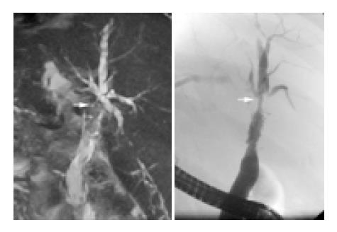
Figure 2 Comparison of ERC and MRC in posttransplant biliary stenosis.
ITBL type III with hilar stenosis (arrow) and multiple peripheral duct stenoses. Left: MRC, right: ERC. The dominant hilar stenosis (arrow) is seen with both methods. The peripheral stenoses are better seen in ERC.
- Citation: Zoepf T, Maldonado-Lopez EJ, Hilgard P, Dechêne A, Malago M, Broelsch CE, Schlaak J, Gerken G. Diagnosis of biliary strictures after liver transplantation: Which is the best tool? World J Gastroenterol 2005; 11(19): 2945-2948
- URL: https://www.wjgnet.com/1007-9327/full/v11/i19/2945.htm
- DOI: https://dx.doi.org/10.3748/wjg.v11.i19.2945


