Copyright
©2005 Baishideng Publishing Group Inc.
World J Gastroenterol. May 21, 2005; 11(19): 2916-2921
Published online May 21, 2005. doi: 10.3748/wjg.v11.i19.2916
Published online May 21, 2005. doi: 10.3748/wjg.v11.i19.2916
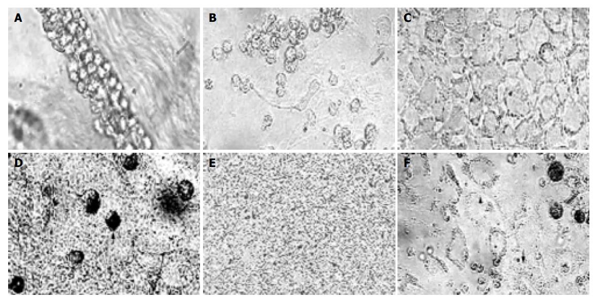
Figure 1 Epithelial cells in primary culture.
A: Ingredients in the sediment after centrifugation (×200); B: colonies formed in cells cultured for 6 d (×100); C: pavement-like cells formed in cells cultured for 9 d (×200); D: immune positive cells (↑) (×400); E: no staining in negative group (×100); F: cell movement, adherent cells (▲) and just adherent cells (↓) (×100).
- Citation: Wang L, Li Q, Duan XL, Chang YZ. Effects of extracellular iron concentration on calcium absorption and relationship between Ca2+ and cell apoptosis in Caco-2 cells. World J Gastroenterol 2005; 11(19): 2916-2921
- URL: https://www.wjgnet.com/1007-9327/full/v11/i19/2916.htm
- DOI: https://dx.doi.org/10.3748/wjg.v11.i19.2916


