Copyright
©2005 Baishideng Publishing Group Inc.
World J Gastroenterol. May 14, 2005; 11(18): 2704-2708
Published online May 14, 2005. doi: 10.3748/wjg.v11.i18.2704
Published online May 14, 2005. doi: 10.3748/wjg.v11.i18.2704
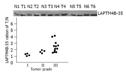
Figure 3 Analysis of the expression of LAPTM4B-35 with the differentiation status of HCCs.
(a) Western blot analysis of LAPTM4B-35 in HCC tumor tissues (T), paired noncancerous liver tissues (N). (Tumor grade I: T3,T5; grade II: T1,T6; grade III: T2,T4 ) (b) Correlation between LAPTM4B-35 protein level and tumor grade. Paired tumor (T) vs adjacent noncancerous liver tissues (N) from 20 HCC patients were compared for their LAPTM4B expression by Western blot. Each spot in the figure represents the ration (T/N) of the LAPTM4B-35 expression (tumor vs adjacent noncancerous liver tissue) from one patient (P<0.05).
- Citation: Peng C, Zhou RL, Shao GZ, Rui JA, Wang SB, Lin M, Zhang S, Gao ZF. Expression of lysosome-associated protein transmembrane 4B-35 in cancer and its correlation with the differentiation status of hepatocellular carcinoma. World J Gastroenterol 2005; 11(18): 2704-2708
- URL: https://www.wjgnet.com/1007-9327/full/v11/i18/2704.htm
- DOI: https://dx.doi.org/10.3748/wjg.v11.i18.2704


