Copyright
©2005 Baishideng Publishing Group Inc.
World J Gastroenterol. May 7, 2005; 11(17): 2616-2625
Published online May 7, 2005. doi: 10.3748/wjg.v11.i17.2616
Published online May 7, 2005. doi: 10.3748/wjg.v11.i17.2616
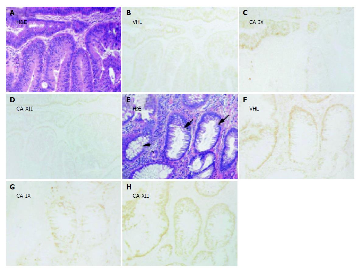
Figure 4 Immunohistochemical staining of pVHL, CA IX and CA XII in parallel sections of (B-D) moderate (lower part) and grave (upper part) adenomas and of (F-H) transitional epithelium (small arrows) located between histologically normal region (arrowhead) and grade I adenocarcinoma (not shown).
Corresponding H&E staining is shown in panels A and E. Original magnifications, ×100.
- Citation: Kivela AJ, Parkkila S, Saarnio J, Karttunen TJ, Kivela J, Parkkila AK, Bartosova M, Mucha V, Novak M, Waheed A, Sly WS, Rajaniemi H, Pastorekova S, Pastorek J. Expression of von Hippel-Lindau tumor suppressor and tumor-associated carbonic anhydrases IX and XII in normal and neoplastic colorectal mucosa. World J Gastroenterol 2005; 11(17): 2616-2625
- URL: https://www.wjgnet.com/1007-9327/full/v11/i17/2616.htm
- DOI: https://dx.doi.org/10.3748/wjg.v11.i17.2616


