Copyright
©2005 Baishideng Publishing Group Inc.
World J Gastroenterol. Apr 28, 2005; 11(16): 2426-2430
Published online Apr 28, 2005. doi: 10.3748/wjg.v11.i16.2426
Published online Apr 28, 2005. doi: 10.3748/wjg.v11.i16.2426
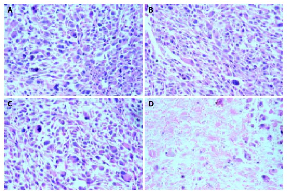
Figure 2 Histological examination of colon cancer xenografts on the 7th d after treatment (hematoxylin and eosin staining).
A: (group B-E-), B: (group B-E+) and C: (group B+E-) contained markedly atypical cells arranged irregularly; D: (group B+E+) exhibited massive necrosis with a moderately intense mononuclear infiltration (40×).
- Citation: Zheng MH, Feng B, Li JW, Lu AG, Wang ML, Hu WG, Sun JY, Hu YY, Ma JJ, Yu BM. Effects and possible anti-tumor immunity of electrochemotherapy with bleomycin on human colon cancer xenografts in nude mice. World J Gastroenterol 2005; 11(16): 2426-2430
- URL: https://www.wjgnet.com/1007-9327/full/v11/i16/2426.htm
- DOI: https://dx.doi.org/10.3748/wjg.v11.i16.2426


